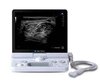
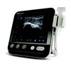
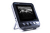
SONIMAGE HS1 Compact Ultrasound System

See more with the SONIMAGE® HS1 Compact Ultrasound System. Delivering superior image quality and needle visualization, you can make patient care decisions with confidence across a variety of point-of-care environments – from the clinic to the bedside.

  • Superior image quality for confident diagnosis
  • Improves accuracy Simple Needle Visualization (SNV®) improved needle visibility
  • Easy to use intuitive touch screen

See more clearly

The HS1 System, with enhanced signal penetration, increased color flow sensitivity, and improved resolution, is capable of detailed tissue differentiation, detecting structures as small as several hundred microns. 

Save time

The UltraAdjust image optimization feature allows you to change imaging parameters by simply adjusting depth. Various imaging parameters such as trapezoid setting, contrast enhancements, SI filter and focus position are automatically optimized “with the touch of the depth button”.

  • Improve your workflow
  • MSK presets linked to anatomical structures are optimized when depth is adjusted

Enjoy the ease of handsfree

Voice activation of the SONIMAGE® HS1 Ultrasound System allows you to operate handsfree during interventional procedures:

  • Simple and accurate voice activation
  • Handsfree system operation
  • No need to break the sterile field
  • Focus more on the patient
  • Enhance diagnostic efficiency

Increasing your comfort, the HS1 System voice control operation features proper operator ergonomics at the point-of-care.

Improves precision

Panoramic view is an imaging process that produces an image that provides both qualitative and quantitative information. Panoramic imaging stitches a series of images to give you one long image in order to:

  • Assess larger lesions
  • Construct a cross-section image of a structure
  • Show the relationship of two structures in a single image

Panoramic imaging technology widens the field of view for precise clinical diagnosis and interventional procedures.

Assess changes easily

Strain Elastography is a real-time qualitative and quantitative imaging software feature that calculates and displays the relative stiffness of tissue. The image is generated from strain generated by uniform pressure of the transducer on the tissue. You can use elastography to visibly assess changes in tendon and ligament elasticity.

Deep and superficial scanning

Unique nanofabrication technology combines both material and machining expertise to produce the SONIMAGE HS1 L18-4 linear transducer. This broad frequency linear probe offers both high sensitivity as well as greater penetration, allowing you to scan both deep and superficial joints and structures.

Access difficult areas

The HL18-4 hockey stick linear transducer for musculoskeletal (MSK) and pain management applications reaches difficult to access areas easily with its small footprint and maneuverability. You can evaluate joints in the fingers and ankle more readily with the probe’s angulation, improved control and greater contact with the anatomy. The high frequency probe provides excellent resolution in the near field for tissue differentiation and visualizes color flow with outstanding Doppler sensitivity.

Increased needle visibility and accuracy

The SONIMAGE HS1 Simple Needle Visualization (SNV®) feature incorporates an advanced algorithm that utilizes both the in-plane and out-of-plane methods to improve needle visibility, especially in steep angle approaches. The resulting clarity of the needle, both tip and shaft, enables increased accuracy in needle placement, making the portable ultrasound system an ideal solution for pain management guided injections.

Get organized

Track and organize ultrasound images efficiently with the SONIMAGE HS1 Rheumatology report package. Still images, along with real time clips, can be stored and assigned to each joint. The RA protocol is integrated and customizable. It can be operated from a foot switch allowing you to move rapidly through the exam without taking your hands from the probe.

Work remotely

From the office, receive remote consultation with Remote Assist. This virtual feature can troubleshoot the system, optimize an image, provide follow up applications support, and offer educational training. And you never have to leave the facility.

More information

  • Blue Moon for Ultrasound Systems
    Brochure
    Blue Moon for Ultrasound Systems Brochure M2105 0323 RevA
  • Simple Needle Visualization
    Brochure
    Simple_Needle_Visualization_v2_M1485_0519_RevBMG2
  • Enhanced Warranties for Ultrasound Systems
    Brochure
    Enhanced Service Coverage for Ultrasound Brochure M2117 0323 RevA
  • Midlands Orthopaedics & Neurosurgery enhances needle-guided imaging with the SONIMAGE® MX1 Portable Ultrasound System
    Case Study / Whitepaper
    Midlands Orthopedics Case Study M2037 0922 RevA
  • Ultrasound Ease of Use Case Study
    Case Study / Whitepaper
    Ultrasound Ease of Use Case Study M2097 0123 RevA

News and Insights

Upcoming events

Get in touch about SONIMAGE HS1 Compact Ultrasound System