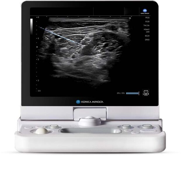
SNV – Simple Needle Visualization
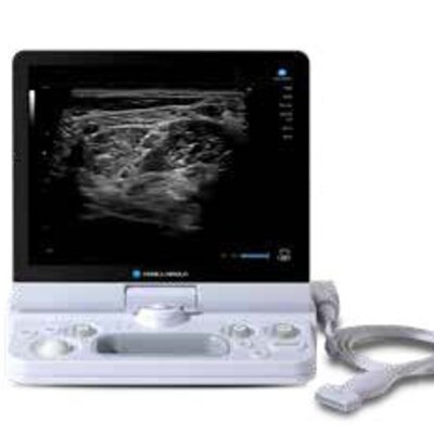
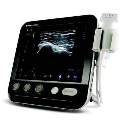
Improve your needle visibility, especially in steep angle approaches, with Konica Minolta’s SNV® software. Incorporating an advanced algorithm that utilizes both the in-plane and out-of-plane methods, the SNV feature increases your accuracy in needle placement, making it the ideal solution for guided injections. SNV is not a stand-alone product and requires either an SONIMAGE® HS1, SONIMAGE® HS2 or an SONIMAGE® MX1 Ultrasound System.
- Enhanced needle visibility, even out-of-plane
- No special needle required
- Increases needle placement accuracy
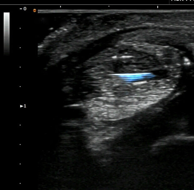
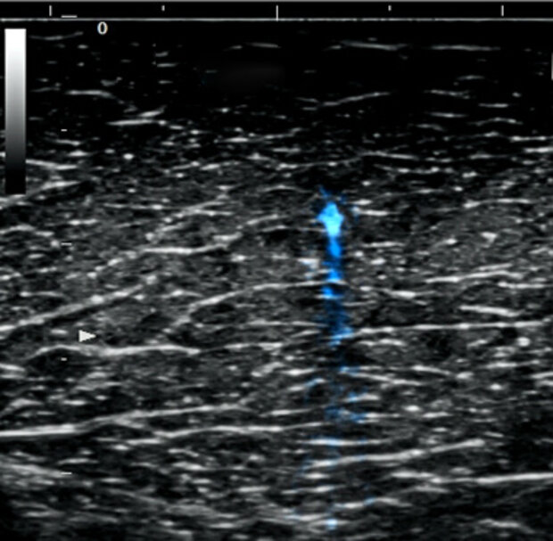
Greater needle visibility
Needle visualization is essential for accurate and successful ultrasound-guided procedures. From regional anesthesia to biologic injections, ultrasound not only images the anatomical structures but also highlights the advancing needle. Simple Needle Visualization (SNV®) software, available with the SONIMAGE® HS2, SONIMAGE® MX1 and SONIMAGE® HS1 Ultrasound Systems, provides greater needle visibility of both the tip and shaft for confident needle placement.
Enhanced needle guidance for interventional procedures:
- Adjust the sensitivity of the needle visualization depending on the type of tissue
- Program prompts help determine the proper level of adjustment
- Auto setting takes the guesswork out of where to steer the ultrasound beam, or work manually by choosing the direction for beam placement
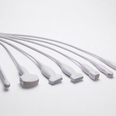
- Can be used with both linear and curved transducers
- Improve injection accuracy by visualizing injectate
