Dynamic Digital Radiography
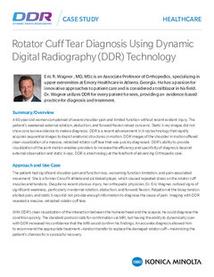
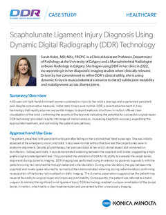
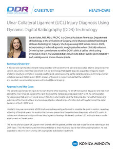
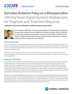
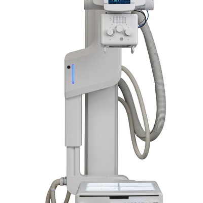
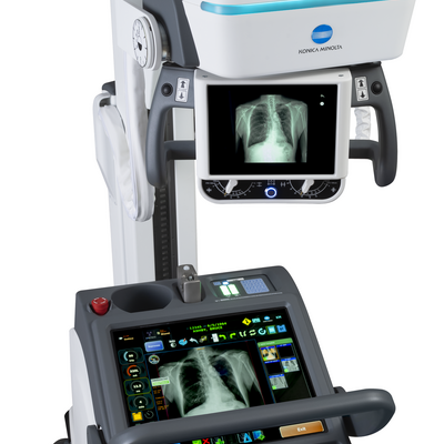
Dynamic Digital Radiography (DDR) allows you to observe movement like never before. This novel low-dose X-ray imaging technique enables visualization of anatomy in motion.
The DDR can acquire up to 15 sequential radiographs per second and play them back as a cine loop, allowing you to observe the physiological cycle and as individual radiographic images (up to 17"x 17" in size). This advancement in digital X-ray technology is FDA-approved and available in many of our systems. DDR is X-ray that moves!
Advantages of X-ray that Moves:
- Fast Exam: In less than a minute, X-ray that Moves gives clinicians up to 20 seconds of physiological movement with a simple acquisition, performed by radiology staff without the need for physician presence
- Low Dose Radiation: With an X-ray that Moves, radiation is lower than in an average fluoroscopy exam.
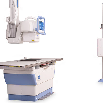
- Convenient & Versatile: The X-ray that Moves system performs all standard X-rays, and images are taken standing, seated or on a table.
See Radiology like never before
Dynamic Digital Radiography (DDR) allows you to observe movement like never before. This novel low-dose X-ray imaging technique enables visualization of anatomy in motion. The DDR can acquire up to 15 sequential radiographs per second, allowing you to observe physiological cycles, as well as individual radiographic images (up to 17"x 17" in size). X-ray that Moves is NOT Fluoroscopy – it is in fact the X-ray precursor to CT or MRI. Cineradiography is X-ray in motion that is derived using digital radiography
- PACS agnostic
- Dose equivalent to about 2 standard X-rays
- Short exposure time for optimal productivity
- Synchronized multi-frame acquisition
- Multi-function X-ray system for dynamic and static images for cost and workflow effectiveness
- Up to 300 images acquired over 20 seconds creates a “cine-loop”
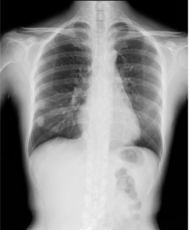
Thorax Images
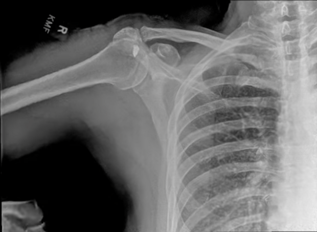
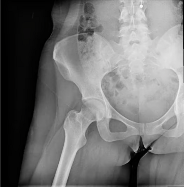
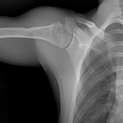
Musculoskeletal applications
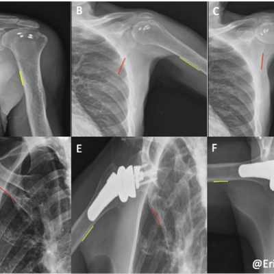
DDR supports diagnosing musculoskeletal conditions by presenting diagnostic detail in full motion.
With DDR, orthopedists can quantify the dynamic relationship between bones and soft tissue through the full range of motion. Having a full view of the musculoskeletal system in motion in weight-bearing or resting positions helps orthopedic specialists provide faster and more detailed diagnoses to improve the quality of care.
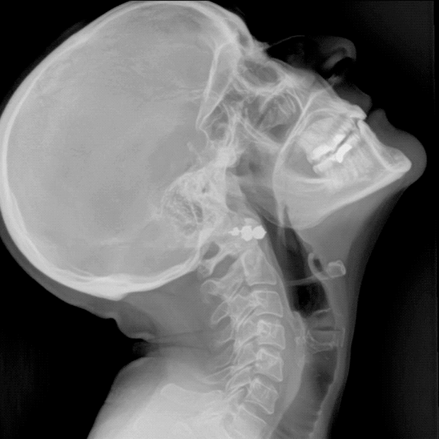
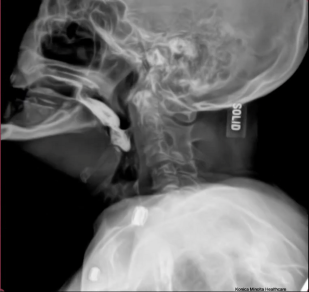
C-Spine
L-Spine
Upper Extremities
Lower Extremities
Swallow Studies
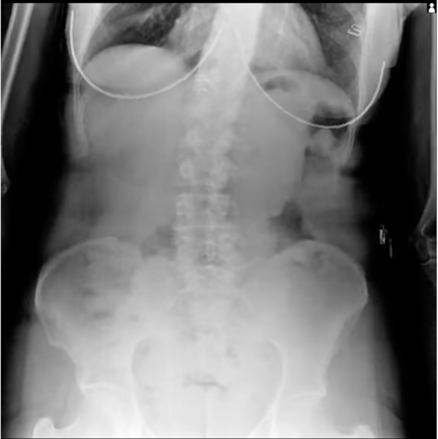
Post Bariatric Sleeve
HSG Series
Increasing Clinical Value
Advantages of X-ray that Moves
-
Fast Exam
In less than a minute, X-ray that Moves gives clinicians up to 20 seconds of physiological movement with a simple acquisition, performed by radiology staff without the need for physician presence
-
Low Dose Radiation
With X-ray that Moves, radiation is lower than an average fluoroscopy exam.
-
Convenient & Versatile
The X-ray that Moves system performs all standard X-rays, and images are taken standing, seated or on a table.
DDR Exams are Used to Assess Spine Stability and Joint Motion
Standard X-ray exams usually show a static view of the span in a flexed and extended positions. DDR can help assess spine stability by providing a detailed view of the full range of motion. Dynamic imaging has been shown to help assess joint and extremities in motion. Many orthopedic facilities are adding a DDR views of spine and extremities to diagnose stability, joint space, assess sources of pain biomechanics, musculoskeletal injury, such as whiplash, as treatment follow-up , and postoperative evaluation of movement before ordering a more advanced exam like CT or MRI. Furthermore, showing patients the joint movement makes communication simpler and more effective.
The experts speak
DDR uses in Pulmonology
DDR uses in Orthopaedics
In the Press
Recent Scientific Papers
-
Noriaki Wada Assessment of pulmonary function in COPD patients using dynamic digital radiography: A novel approach utilizing lung signal intensity changes during forced breathing. European Journal of Radiology
-
Yuzo Yamasaki Diagnostic accuracy and added value of dynamic chest radiography in detecting pulmonary embolism: A retrospective study. European Journal of Radiology Open
-
Ziyang Xia Investigation of diaphragmatic motion and projected lung area in diaphragm paralysis patients using dynamic chest radiography. Quantitative Imaging in Medicine and Surgery
