Assessing C-spine Injuries with Dynamic Digital Radiography

Injuries to the cervical spine (C-spine) are often the most severe type of spinal cord injuries that can cause permanent changes in strength, sensation and other functions, including paralysis and death.
Featuring Dr. Neill Wright, Blessing Health
Injuries to the cervical spine (C-spine) are often the most severe type of spinal cord injuries that can cause permanent changes in strength, sensation and other functions, including paralysis and death. Trauma is the most common cause of cervical spine injuries; non-traumatic causes include compression fractures and inflammation.Diagnostic imaging, such as X-ray, CT and MRI, is typically utilized in the evaluation of C-spine injuries. However, inaccurate assessment and diagnosis of cervical spine injuries remain a common issue in trauma medicine, with estimates of patients receiving a delayed diagnosis between 5-20%1,2.“One of the challenges of evaluating spinal disorders, especially in the C-spine, is how the spine works in motion,” explains Neill Wright, MD, neurosurgeon, Blessing Health System (Quincy, IL). Dr. Wright is an internationally renowned cervical spine expert who has performed more than 2,000 C-spine surgeries and holds several patents related to C-spine procedures, including developing the C2 laminar fixation technique for the treatment of atlantoaxial instability.
“Most of the imaging we do is with the patient lying supine, such as during a CT or MRI study. That doesn’t allow for assessment of some of the changes that gravity may have on the spine, and it doesn’t help us assess instabilities or ligamentous laxity of the spine,” says Dr. Wright. While CT and MRI are commonly utilized for evaluating C-spine injuries, X-ray, or digital radiography (DR), is also used to capture images of the patient standing, as well as flexion and extension.
Additionally, the clinician can only visualize the forward and backward position of the neck, not side-to-side. According to Dr. Wright, there are other forms of instability in the cervical area requiring surgical intervention that might not be visible with this type of imaging.
To obtain as much information as possible from the radiographs, Dr. Wright worked with a radiologist to create an imaging protocol to capture the lateral movement of the C-spine. Patients are imaged with X-rays while in an open-mouth position to visualize C1 and C2, as well as with right and left lateral motion of their head. Due to the complex anatomy and projectional variation of these radiographs, they can be difficult to interpret and distinguish normal C-spine anatomy from injury or pathology3.
There are also limitations to static flexion and extension imaging, including an inability to view or assess what is happening with the patient’s cervical spine while in motion.
A new dynamic imaging modality
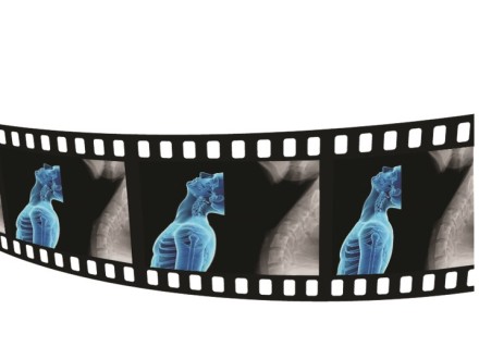
Dynamic Digital Radiography (DDR) from Konica Minolta Healthcare is an FDA-cleared, award-winning enhanced X-ray technology that provides a series of individual digital images acquired at high speed and low dose. The resulting cineloop enables clinicians to observe the motion of anatomical structures over time, improving diagnostic capabilities. DDR is available on Konica Minolta’s KDR® Advanced U-Arm, a compact and efficient DR system that supports imaging views commonly required in MSK and radiology, including wheelchair and table work.
DDR can help assess spine stability4 by providing a detailed view of the full range of motion in both front to back and side to side views. Assessing joints and extremities in motion has been shown to increase diagnostic capabilities5.
DDR gives me a much better picture of the patient’s instability...
“DDR gives me a much better picture of the patient’s instability compared to what I could see on previous ‘dynamic’ films,” explains Dr. Wright. Prior to DDR, patients with suspected C1 instability could be referred for a digital motion X-ray (DMX) with a chiropractor. However, DMX is a fluoroscopic imaging device, delivering a much higher dose than X-ray and is not readily available. By comparison, DDR delivers a dose that is marginally higher than a traditional static C-spine series.
“My real excitement about the DDR technology is looking at C1 and C2 instability with that dynamic movement. I'm starting to see patients who are complaining of significant neck pain with a sense of instability. This is where all traditional imaging has failed to show any problems. However, with DDR and the patient performing a dynamic lateral bend, we're seeing instability or laxity at C1 and C2 that was not otherwise diagnosed.”
Now, any patient receiving a cervical consult receives a DDR exam with a protocol designed by Dr. Wright at the new Blessing Health Center 4800 Maine. The technologist first captures a lateral view with full extension and full flexion. Then, an AP static image and an AP dynamic series of images are captured. The dynamic series includes an open-mouth view with lateral bending, which involves the patient tilting their head all the way to the right and then all the way to the left. The dynamic exam is performed with low dose, nearly equivalent to the static X-ray views taken for each patient.
“It is seamless, cost-effective and now it is part of our normal radiographic workup.”
The DDR exam is also easier for the technologists to obtain, as they can directly visualize the exam as the patient is moving on their workstation. If needed, they can quickly make adjustments to the patient’s position to ensure the correct views are captured, reducing the need to repeat an exam or extending the length of the exam by taking multiple additional views.
“This technology very much filled an unmet need, and so it was immediately adopted in our center,” Dr. Wright adds. “It is seamless, cost-effective and now it is part of our normal radiographic workup. I could immediately see the potential of this technology, so having it in our center is a winner on all fronts.”
References
- Goradia D, Blackmore CC, Talner LB, Bittles M, Meshberg E. Predicting radiology resident errors in diagnosis of cervical spine fractures. Acad Radiol. 2005 Jul;12(7):888-93.
- Platzer P, Hauswirth N, Jaindl M, Chatwani S, Vecsei V, Gaebler C. Delayed or missed diagnosis of cervical spine injuries. J Trauma. 2006 Jul;61(1):150-5.
- Matthew R. Skalski, DC, DACBR George R. Matcuk, Jr, MD Wende N. Gibbs, MD The Art of Interpreting Cervical Spine Radiographs. RadioGraphics 2019; 39:820–821 https://doi.org/10.1148/rg.2019180148
- Wright, Neill M., and Carl Lauryssen. "Vertebral artery injury in C1-2 transarticular screw fixation: results of a survey of the AANS/CNS section on disorders of the spine and peripheral nerves." Neurosurgical Focus 4.2 (1998): E2.
- George S.I. Sulkers, Niels W.L. Schep, Mario Maas, Simon D. Strackee, Intraobserver and Interobserver Variability in Diagnosing Scapholunate Dissociation by Cineradiography, The Journal of Hand Surgery, Volume 39, Issue 6, 2014, Pages 1050-1054.e3, ISSN 0363-5023, https://doi.org/10.1016/j.jhsa.2014.03.014.
