
University of Alabama at Birmingham Reaches 1,000 Patient Pulmonary Exams With Dynamic Digital Radiography

The University of Alabama at Birmingham (UAB) Hospital achieves the significant milestone of imaging more than 1,000 patients with Dynamic Digital Radiography (DDR) technology since it was installed in June 2021
Wayne, NJ, March 7, 2023 – Konica Minolta Healthcare Americas, Inc., congratulates the University of Alabama at Birmingham (UAB) Hospital for achieving the significant milestone of imaging more than 1,000 patients with Dynamic Digital Radiography (DDR) technology since it was installed in June 2021. DDR has been utilized in the Kirklin Clinic of UAB Hospital for pulmonary imaging in pre and post lung transplant, post heart transplant, after COVID infection, chronic obstructive pulmonary disorder (COPD) and lung cancer patients, as well as for evaluating diaphragm dysfunction. UAB Medicine is a leading lung transplant center and one of busiest in the US, with one-year patient and graft survival rates higher than national averages.
DDR is a radiographic technique that provides a series of individual digital X-ray images acquired at high speed and low radiation dose. These images provide diagnostic-quality views of anatomical structures in motion and the ability to visualize the dynamic interaction with physiological changes over time.
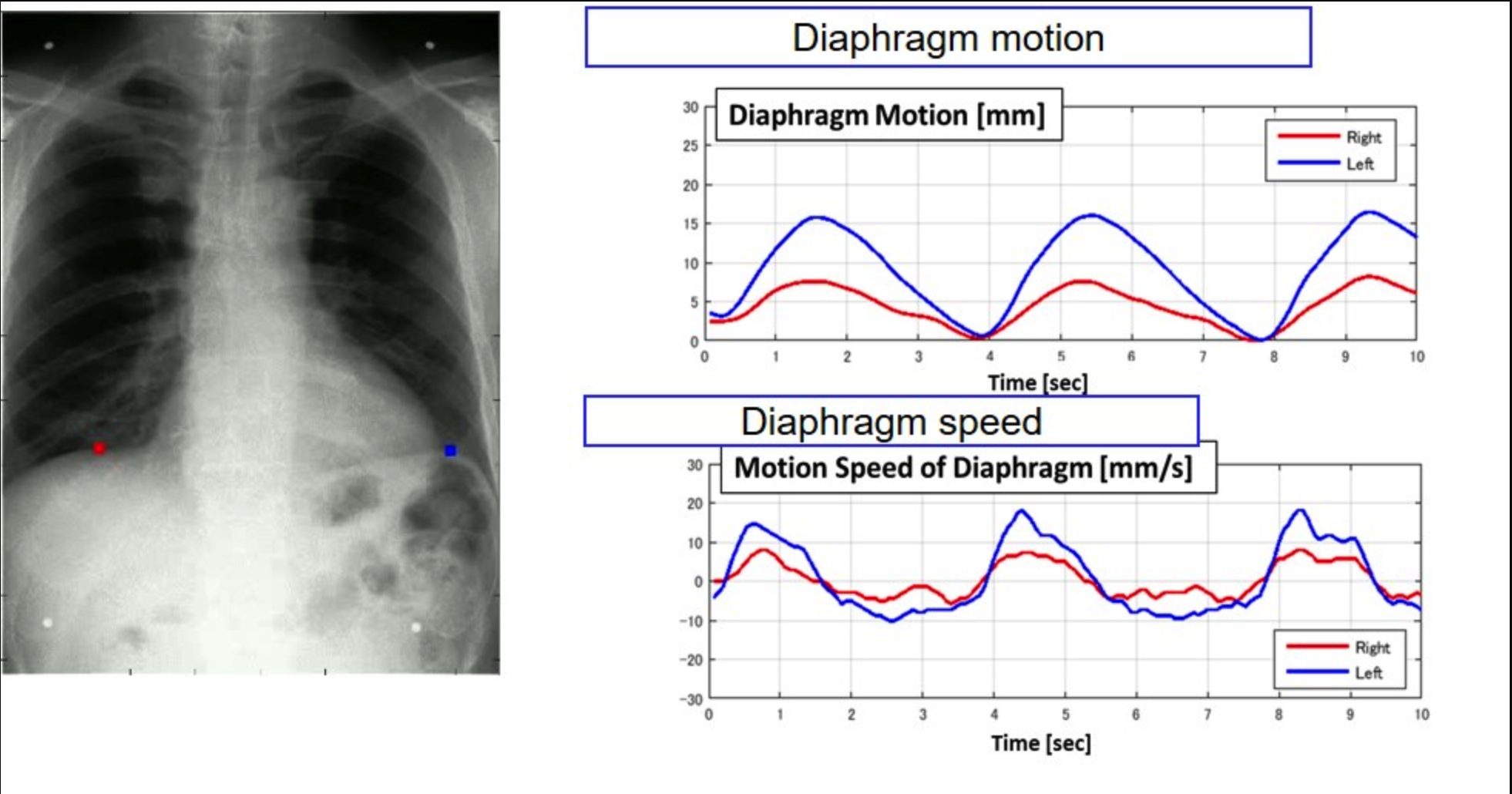
Satinder Singh, MD, FCCP, FNASCI, FSABI, Professor of Radiology & Medicine and Chief of Cardiopulmonary Radiology in the Department of Radiology at UAB, explains that “DDR is a viable replacement for chest fluoroscopy in the evaluation of diaphragm function. It provides good visibility of lung abnormalities and increases our diagnostic confidence. Based on our extensive evaluation and utilization of this technology, we have now transitioned to DDR instead of traditional chest fluoroscopy to evaluate an elevated diaphragm. All lung transplant patients currently receive a DDR exam as part of their pre-surgical workup.”
DDR provides enriched radiographic information and enhanced diagnostic capabilities. Compared to fluoroscopy, the patient’s radiation dose is lower with DDR, comparable to a standard two-view X-ray exam. Dr. Singh and his colleagues at UAB use DDR to routinely assess diaphragm function in transplant patients and, in some cases, monitor treatment effects, examine airways and evaluate pulmonary function in their patients.
At a Konica Minolta Healthcare sponsored Lunch and Learn event held during the Radiological Society of North America (RSNA) 2022 Annual Meeting, Dr. Singh presented initial results from a pilot study led by Donald G. Benson, Jr., MD, PhD, Assistant Professor, Cardiopulmonary Imaging Section, comparing lung perfusion results from DDR with nuclear medicine scintigraphy. The pilot study compared differential lung perfusion between the right and left lungs from both modalities. Results are being analyzed for statistical correlation, including the percent of perfusion in each lung and in different portions of the lung in each patient.
Dr. Benson says, “We are finding out there is no discrepancy between these two modalities in how we manage patients clinically. Our transplant patients already receive a chest X-ray, so by adding DDR to this technique we can get that diaphragm information and perfusion all in one exam, with low radiation dose.”
DDR images are acquired in the same X-ray system and in the study as static images, making this new technology easy to integrate into our current workflow.
Jeff McGough, Senior Director Imaging Services, Department of Radiology, UAB Hospital
“As with any imaging modality, DDR integrates with our HIS and RIS, so the DDR data is automatically moved to our PACS for processing,” says Jeff McGough, Senior Director Imaging Services, Department of Radiology, UAB Hospital. “DDR images are acquired in the same X-ray system and in the study as static images, making this new technology easy to integrate into our current workflow. Additionally, most of our patients are already scheduled to receive a chest X-ray, so adding DDR to the study gives us additional diaphragm information in one exam, with low radiation dose and without creating discomfort for our patients.”
According to McGough, since DDR is a radiographic procedure, acquisition can occur without having a physician present, unlike a fluoroscopic exam. This significantly improves the workflow efficiency for the department. DDR enables efficient routine dynamic imaging, for example sniff tests of their transplant patients, without dedicated scheduling or disrupting the regular reading schedule.
At UAB, DDR is installed on a Shimadzu RADspeed Pro X-ray system, which is also used for conventional static X-ray acquisitions. DDR is also available on Konica Minolta’s KDR® Advanced U-Arm System, KDR® Flex Overhead X-ray System, mKDR xPress™ Mobile X-ray System and the Chiropractic Straight Arm from 20/20 Imaging, a division of Konica Minolta Healthcare.
“Konica Minolta congratulates UAB on achieving this significant milestone of 1,000-plus patient exams using DDR in lung patients,” says John Sabol, PhD, Clinical Research Manager, Konica Minolta Healthcare. “The clinical evidence supporting the use of DDR in pulmonary imaging at UAB is impressive and growing, further demonstrating the clinical value of this novel technology across an array of lung diseases. Drs. Singh and Benson are highly regarded experts in cardiopulmonary imaging, and we look forward to continuing to collaborate with them and the entire clinical team at UAB as they investigate and demonstrate the utility of DDR.”
About Konica Minolta Healthcare Americas, Inc.
Konica Minolta Healthcare is a world-class provider and market leader in medical diagnostic imaging and healthcare information technology. The company’s focus is to contribute to life changing advances through the transformation of primary imaging, allowing the invisible to be seen. Primary imaging, the most commonly used medical imaging technologies, include X-ray, ultrasound and imaging management systems. By advancing these readily available technologies, we can bring greater diagnostic capabilities to the greatest number of people.
With nearly 150 years of endless innovation, imaging is in Konica Minolta’s DNA. From roots as a camera and film manufacturer, the company has cultivated its own technologies and continues to evolve techniques for visualizing what is not visible. Innovation allows the company to be a strong strategic partner, understanding what value means to customers and how Konica Minolta’s innovations can address specific needs and lead to better decisions, sooner.
Konica Minolta Healthcare Americas, Inc., headquartered in Wayne, NJ, is a division of Konica Minolta, Inc. For more information on Konica Minolta Healthcare Americas, Inc., follow us on LinkedIn, Twitter and Facebook, or visit https://healthcare.konicaminolta.us.
Media Contact:
Mary Beth Massat
Massat Media
224.578.2388
https://healthcare.konicaminolta.us
