
Radiological Society of North America Annual Meeting (RSNA) 2025

Imaging the Individual. At the Frontline of Care.

From tailored X-ray, Imaging IT, and Service Solutions to breakthrough innovations, Konica Minolta Healthcare empowers radiology professionals like YOU to further enhance patient outcomes and streamline workflows - so YOU can do more with less. Connect with us at booth 2165 to learn and get inspired. Something extraordinary awaits!
Exhibitor Listing Online Showroom


For more information or to schedule a demo at RSNA complete this form
Events and Presentations
Portable DDR: Bringing Dynamic Chest Imaging to the Bedside
- Presented by: Michaela Cellina, Rossella Catona, MD, Maurizio Ce, MD,BA, Gabriele Drago, MD, Stefano Pedrini, Matilde Pavan, MD, Giancarlo Oliva
- Session Start: 11/30/2025 7:30:00 AM
Teaching Points: Portable DDR enables bedside dynamic chest imaging, offering functional insights into diaphragmatic motion, lung perfusion (contrast-free), and post-procedural complication.
Table of Contents/Outline: DDR is a novel high-resolution imaging technique employing pulsed X-ray emission to rapidly acquire multi-frame sequential images of a targeted anatomical region, rendered as cine loops. Initially developed for orthostatic imaging, its recent integration into portable systems now enables dynamic acquisitions at the patient's bedside, unlocking the potential for new diagnostic insights.
Dynamic Chest Radiography Improves Detection And Diagnostic Confidence Of Lung Cancer Lesions Compared To Conventional Radiography
- Presented by: Koji Takumi, MD, PhD, Ryota Nakanosono, MD, Hiroaki Nagano, MD, PhD, Tsubasa Nakano, Fumitaka Ejima, Masanori Nakajo, MD, Kiyohisa Kamimura, MD, PhD, Takashi Yoshiura, MD, PhD
- Session Start: 12/1/2025 9:30:00 AM
Purpose: To investigate whether dynamic chest radiography (DCR) improves the diagnostic accuracy of conventional chest radiography (CCR) for detecting lung nodules.
Methods and Materials: The study included 100 surgically treated lung cancer lesions. All patients underwent preoperative CCR, DCR, and CT examinations. DCR was obtained using a dynamic flat-panel detector imaging system, equipped with the X-ray moving image analysis workstation (KINOSIS, Konica Minolta, Inc., Tokyo, Japan). The diagnostic confidence for the lesions was evaluated by three chest radiologists on a 3-point scale (2: definite presence, 1: probable presence, 0: difficult to identify) for both CCR and DCR, with a score of 1 or higher defined as a detectable lesion. Artificial intelligence (AI)-based software (CXR-AID, Fujifilm, Tokyo, Japan) also evaluated lesions on conventional chest radiographs.
From Static To Dynamic - Advancing PTE Diagnosis Through The Lens Of DDR
- Presented by: Robin Philip, MBBS, Priscilla Joshi, MBBS, MD
- Session Start: 12/2/2025 12:45:00 PM
Purpose: To evaluate diagnostic accuracy of Dynamic Digital Radiography (DDR) in suspected acute pulmonary thrombo-embolism (PTE) and its potential to overcome the limitations of conventional CT pulmonary angiography (CTA).To evaluate pulmonary perfusion on follow up DDR.To correlate initial and follow up DDR findings with Transthoracic 2D echocardiography (TTE) findings.
Methods and Materials: This prospective study evaluated diagnostic performance of DDR in adult patients with suspected PTE undergoing CTA. It was an interim analysis of 62 patients , 27(43.5 %) were females and 35(56.5 %) were males. TTE was done for patients who presented with acute onset breathlessness. Those with a suspicion of PTE were sent to radiology department for diagnostic CTA.
Automated Ventilatory Function Assessment Using Deep Learning Analysis Of Dynamic Chest Radiography: An Imaging-Based Alternative To Spirometry
- Presented by: Yaya G, Ming Jia, Jiefang Wu, Genggeng Qin, MD, PhD, Weiguo Chen
- Session Start: 12/2/2025 12:45:00 PM
Purpose: To develop an automatic analysis pipeline for ventilatory function assessment using deep learning analysis of dynamic chest radiographys (DCR).
Methods and Materials: We prospectively enrolled 435 patients who underwent DCR and spirometry between August 2022 and December 2024. A deep learning-based cascade system, which comprises two modules (segmentation and classification), was developed to evaluate whether subjects exhibit ventilatory dysfunction and assess the severity of ventilatory dysfunction using DCR. The performance of the models was evaluated using the mean area under the receiver operating characteristic curve (AUC), accuracy, sensitivity, precision, and specificity.
Pulmonary Blood Flow Analysis Using Oblique Dynamic Chest Radiography
- Presented by: Tasuku Nakajima, Ryuichi USHIJIMA, Rie Tanaka, PhD, Aki Kido, MD, PhD, Kyo Noguchi, MD
- Session Start: 11/30/2025 7:30:00 AM
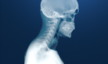
Teaching Points: Dynamic chest radiography (DCR) is a newly developed functional X-ray imaging technique that visualizes pulmonary blood flow without using contrast media (Fig. 1). DCR-based pulmonary blood flow findings are reportedly consistent with those in other imaging modalities, such as lung scintigraphy, pulmonary arteriography, and contrast-enhanced chest computed tomography (CT). However, DCR is usually performed in the posteroanterior (PA) direction, therefore, it cannot analyze the lung regions behind the heart in the PA direction. One solution is oblique imaging, as is done in lung scintigraphy. The purpose of this exhibit is to expose radiologists to a series of clinical cases in order to utilize oblique DCR to assess pulmonary blood flow in daily clinical settings.
Effort-Independent, Non-Contrast Ventilatory Assessment Of Interstitial Lung Disease Progression Using Resting-State Dynamic Chest Radiography
- Presented by: Kojiro Ono, Takefumi Nikaido, Ryoichi Watanabe, Akinori Tsunomori, Yoshinori Tanino, Yoko Shibata, Tsutomu Yoneyama
- Session Start: 11/30/2025 9:30:00 AM
Purpose: This study aimed to evaluate an effort-independent, non-contrast method using resting-state dynamic chest radiography (DCR). Our scheme, Resting-State Silhouette Tracking for Thoracic Respiratory Assessment and Characterization of Kinetics (REST-TRACK), tracks respiratory motion via silhouettes and derives four ventilatory indices based on lung area and diaphragmatic displacement to distinguish healthy lungs from interstitial lung disease (ILD) and to monitor its progression.
Methods and Materials: This IRB-approved retrospective study included 18 ILD patients (≥6 months apart DCR) and 8 healthy controls with no smoking history. DCR was performed in the supine position during resting breathing (15 fps; entrance surface dose of ~1.8 mGy). Respiratory motion was extracted using REST-TRACK, which applies template-matching and deep learning models for silhouette segmentation and tracking.
Dynamic Digital Radiography-Derived Perfusion Indices Reflect Structural And Functional Impairment In COPD With Emphysema
- Presented by: Masahiro Okada, MD, PhD, Kojiro Ono, Takashi Oki, Yasuhiro Gon
- Session Start: 12/4/2025 11:00:00 AM
Purpose: Pulmonary emphysema in chronic obstructive pulmonary disease (COPD) leads to reduced alveolar perfusion and impaired gas exchange. While high-resolution CT (HRCT) quantifies structural changes, non-invasive functional assessment of perfusion remains limited. We aimed to evaluate whether perfusion imaging using dynamic digital radiography (DDR) could serve as a quantitative biomarker for emphysema severity.
Methods and Materials: This prospective single-center study enrolled 40 patients with stable COPD who underwent DDR, HRCT, and spirometry. From breath-hold DDR sequences, pulmonary perfusion maps were generated, and low signal area (LSA) ratios were calculated based on histogram thresholds (5%, 10%, 15%). CT-derived low attenuation volume (%LAV) and Goddard scores were used as reference structural indices.

Dynamic Digital Radiography has received an Innovative Technology designation from Vizient.®
Discover how this novel technology allows you to visualize anatomy in motion.
Learn More About Our Imaging Solutions
Digital Radiography
Creating a faster path to diagnosis and treatment for clinicians and their patients with Dynamic X-ray
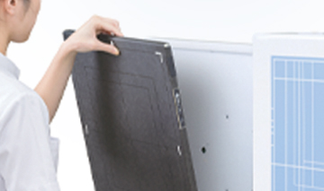
Digital Radiography Detectors
Enhancing performance and efficiency with industry-leading Digital Detectors

Imaging IT
Providing fast access to care from any device, anywhere, with Imaging IT in a cloud framework

Service Solutions
Simplifying the management of your Radiology departments with a comprehensive analytics-driven Productivity Dashboard
